iFS® Advanced Femtosecond Laser
iFS® Advanced Femtosecond Laser
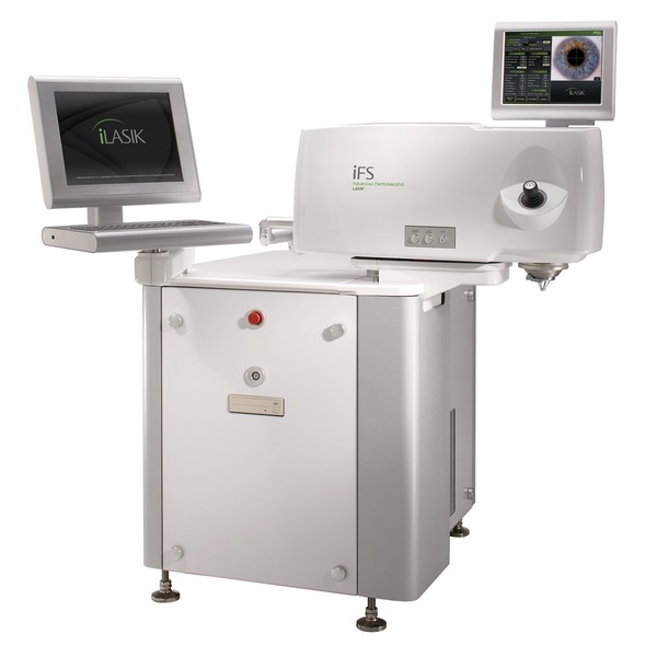
This laser is the only 5th-generation laser installed in the south of the United States, Puerto Rico and the Caribbean. It uses the most advanced technology approved by the FDA to create the needed cutout for the insertion of the KamraInlay used in the surgery to correct presbyopia.
The IFS® Advance Femtosecond Laser uses innovative technology to create a bladeless thin cut of your cornea, allowing you to enjoy an all laser vision correction. This cut is unique to the particular shape of your eye, achieving a better and more precise application of laser pulses, fewer complications and faster visual recovery.
Allegretto Wavelight® EX500 Excimer Laser
Allegretto Wavelight® EX500 Excimer Laser
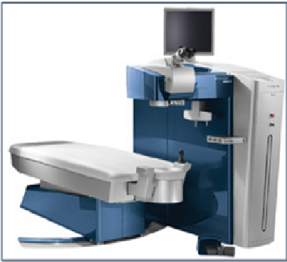
The Food and Drug Administration approved the use of the Allegretto Wavelight® EX500 Excimer Laser for cutting the surface of the cornea in preparation for Lasik refractive surgery. This instrument uses an infrared laser that has the ability to track the eye movement, thus avoiding the refractive errors that occur in surgeries performed without this capability.
With the Allegretto Wavelight we perform a bladeless cut that is strategically positioned at the indicated depth to achieve greater visual acuity in a high precision, fast, painless and individualized procedure.
Femto Laser Lensx
Femto Laser Lensx

ALCON’s LenSx Laser raises the refractive surgery to new levels of precision, specifically in the creation of the required cuts for inserting intraocular multifocal lenses in cataract surgeries.
This laser is designed to make primary and secondary incisions specifically tailored to the contours of the patient's cornea, ensuring maximum accuracy in the surgery and the best visual acuity that your eye can get.
True Vision 3D Guide Systems
True Vision 3D Guide Systems
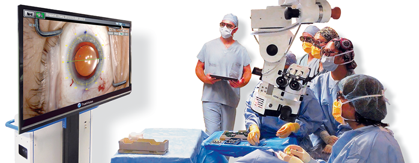
TrueGuide® is a tri-dimensional assisted surgery system, specially designed for cataract surgery and refractive visual correction procedures. TrueGuide features specifically designed surgical parameters for each patient, allowing precise positioning of the intraocular lens, thus improving the refractive surgery results. Instituto de Ojos is the only ophthalmologic facility in Puerto Rico and the Caribbean with this advanced technology to optimize and individualize refractive cataract surgeries, achieving greater precision in the surgery.
Sistema VERION™
Sistema VERION™
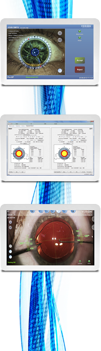
This system is used in cataract surgeries to maximize and optimize the accuracy of the incision and intraocular lens alignment using as a guide the patient's eye images in high resolution. VERION™ centralizes and automatically aligns both, monofocal and multifocal lenses.
VERION’s reference unit also allows to determine the most appropriate intraocular lens to achieve the desired visual correction.
OCT
Optical Coherence Tomography (OCT)
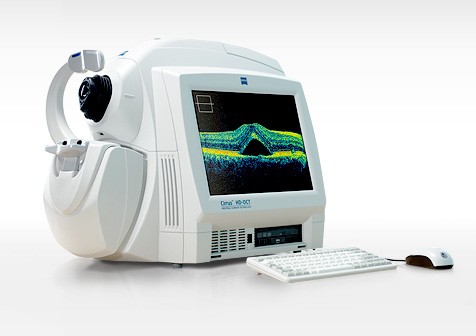
Optical Coherence Tomography (OCT) is an exploration technique, useful for retina and optic nerve studies. Its main feature is the possibility of obtaining high-resolution images of the surface of the retina and its layers.
For more information on this technology please visit the manufacturer's website.
Humphrey: HFA II series i
Humphrey: HFA II series i
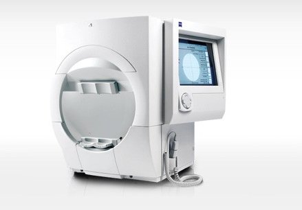
Analysis and advanced diagnostics.
- Guided Progression Analysis™ (GPA™): this perimetry progression analysis software automatically identifies statistically significant changes.
- Visual Field Index™ (VFI™): a simple and intuitive global index that determines the percentage of visual loss of all visual fields.
- STATPAC™: the language of perimetry compares the results with normative databases by age and glaucoma databases.
- SITA™: patented acquisition algorithm for fast and accurate measurements of the visual field threshold; the most commonly used test strategy normally incorporates patient responses in real time.
Si desea más información sobre esta tecnología puede visitar la página del fabricante.
EasyScan
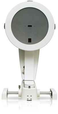
EasyScan
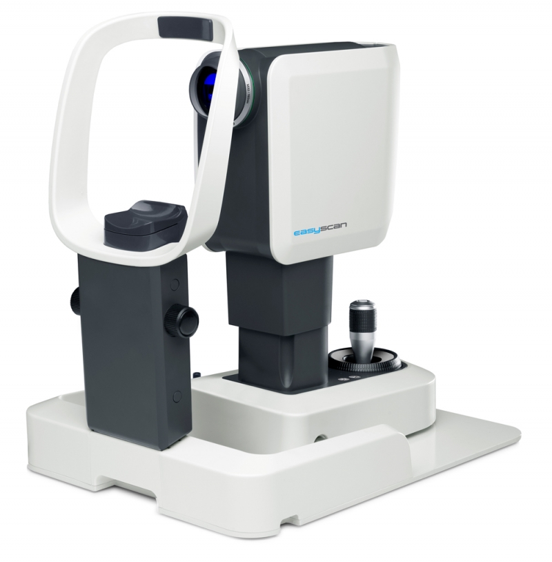
Used for early diagnosis of retinal conditions such as diabetic retinopathy, macular degeneration and age-related glaucoma.
Provides higher contrast than a traditional camera background.
Has better penetration of opacities such as cataracts.
For more information on this technology please visit the manufacturer's website.
Pentacam
Pentacam
Pentacam is a camera with a rotation system based on Scheimpflug for the anterior segments analysis. It captures the thickness of the cornea and takes data from topography and elevation of the anterior and posterior face of the cornea. The basic software includes maps previews, fast screening reports, color map, 4 refraction maps, overview of Scheimpflug, general images, virtual eye, tomography, image of the iris, topometric and comparison screens images.
The OCULUS - Pentacam® software can be customized and updated anytime, by adding an optional module adapted to your needs.
For more information on this technology visit the manufacturer's website.
AcuTarget HD
AcuTarget HD
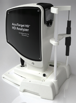
El AcuTarget HD es la herramienta de diagnóstico que ayuda a los médicos para ver realmente lo que el paciente está viendo, mide con precisión la profundidad de foco, la calidad visual, la película lagrimal y puntos de referencia oculares únicas para ayudar a los médicos a seleccionar la mejor opción de tratamiento para un amplio espectro de pacientes incluidos presbicia, LASIK, cataratas y ojo seco.
Si desea más información sobre esta tecnología puede visitar la página del fabricante.
California
California
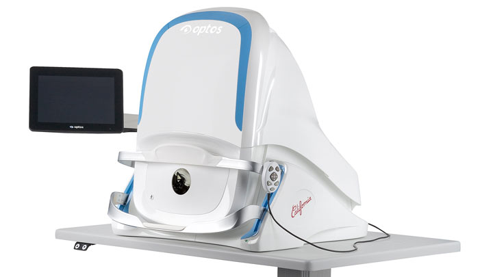
With California, Optos incorporates new hardware and software technology that allows to see more, discover more and treat more effectively most eye diseases, thus improving the health of patients. We are committed to further strengthen the clinical evidence and demonstrate the importance of diagnostic techniques for imaging the entire retina.
California includes a new diagnostic imaging modality UWF optomap®, the indocyanine green angiography (iga), which maintains:
- Compound color
- Free of red
- Auto fluorescence (af)
- Fluorescein angiography (fa)
The new patented optical hardware optimizes and maintains Optomap® resolution of images during scanning of the retina with greater clearly in the far periphery.
The image overlay allows comparison of color images and free of red, af images, fa or iga. Furthermore, comparisons between different images or different dates can be made by scrolling through the stored images.
In addition to the advantages provided by all Optos UWF devices, such as 200 degrees’ panoramic views or capture of up to 82% of the retina in a single image in multiple modalities, and that professionals can see more than 50% of the retina in comparison with conventional diagnostic devices, California offers the following advantages:
- Diagnostic technique through the high resolution image without mydriasis through several 2 mm cataracts or pupils to save time in crowded consultations.
- Review of images based on search engine allowing integration and access to data from any computer or tablet.
- Interlaced acquisition angiography allows to capture parallel images fa and iga without having to switch manually between modes of imaging.
If you would like more information about this technology you can visit the manufacturer's website.